
For most people, radiation and heart treatment exist in entirely separate mental categories. Radiation is for tumors. The heart is treated with drugs, stents, and surgeons threading catheters through blood vessels. That clean division, however, is quietly beginning to blur – and a study published in April 2026 may represent one of the more significant moments in that shift.
The patients at the center of this story had already exhausted most of what modern cardiology could offer them. They had undergone multiple invasive procedures. They had tried medication after medication. Their hearts kept misfiring in ways that were not just uncomfortable but potentially fatal. And yet, after a single, noninvasive treatment session that left no surgical scars and required no anesthesia, something notable happened: their dangerous heart rhythm episodes dropped by nearly 80%.
The treatment was proton beam therapy, a technology borrowed from oncology. The condition it targeted was ventricular tachycardia, one of the most serious rhythm disorders the heart can produce. What researchers found in this small but carefully documented first-in-human trial has prompted cardiologists and radiation oncologists alike to sit up and take notice.
What Is Ventricular Tachycardia – and Why Does It Matter So Much?
Ventricular tachycardia (VT) is defined as three or more consecutive heartbeats at a rate above 100 beats per minute, arising from the ventricles – the heart’s lower pumping chambers. It is a potentially life-threatening arrhythmia and is responsible for the majority of sudden cardiac deaths in the United States.
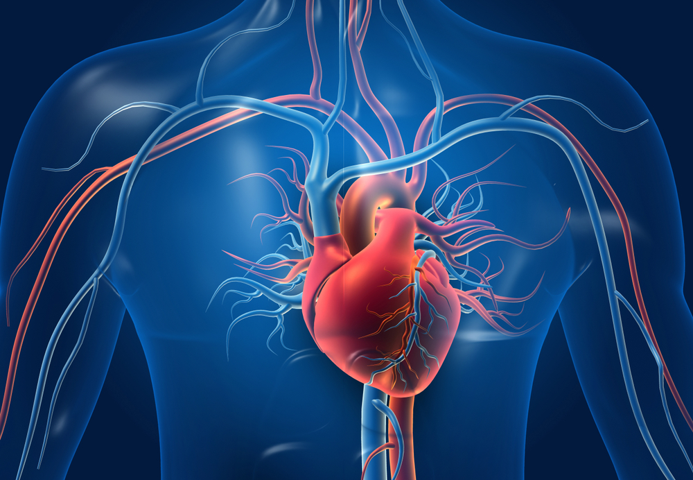
Understanding why it happens requires a brief look at cardiac anatomy. Ventricular tachycardia is commonly caused by scar tissue within the heart’s pumping chambers. That scar tissue behaves like a short electrical circuit: when the right trigger comes along, it fires repeatedly around the circuit, producing rapid, chaotic heartbeats. In most cases, sustained monomorphic VT – the most common and dangerous variety – results from scar tissue caused by a prior heart attack or cardiomyopathy (disease of the heart muscle itself).
The scale of the problem is substantial. Sudden cardiac death, defined as an unexpected death from cardiovascular causes within one hour of symptom onset, accounts for an estimated 10% to 15% of all deaths globally, and more than 356,000 individuals experience out-of-hospital cardiac arrests in the United States annually. Ventricular tachycardia is a major contributor to sudden cardiac death, particularly in patients with ischemic and nonischemic cardiomyopathy, where its degeneration into ventricular fibrillation can result in cardiac arrest.
The Treatment Gap: When Standard Options Run Out
Cardiologists currently have three main tools for managing ventricular tachycardia: antiarrhythmic medications, implantable cardioverter-defibrillators (ICDs, devices surgically implanted in the chest that deliver shocks when VT is detected), and catheter ablation.
Catheter ablation is typically the most aggressive of these approaches. In this invasive procedure, doctors thread thin, flexible wires – catheters – through the patient’s blood vessels to reach the heart, then use heat or cold energy to create precise scars that block the abnormal electrical signals. When it works, it can be highly effective. In patients with structural heart disease, catheter ablation achieves a 70% freedom from VT recurrence at one year. That means roughly 30% of patients see their arrhythmia return within a year despite seemingly successful procedures.
For those who relapse, the options narrow significantly. Catheter ablation of VT is associated with complications in 6% to 7% of cases, including vascular access complications, cardiac tamponade, and stroke, and a 30-day all-cause mortality rate of 4% to 5%. Patients with multiple failed ablations and advanced heart disease – especially those with significantly reduced heart pumping function, known as reduced ejection fraction (the percentage of blood the heart pumps out with each beat) – represent a group that medicine has historically struggled to help.
Implantable cardioverter-defibrillators are the primary management strategy for VT and reduce the incidence of death, but they do not reduce VT recurrences. High VT burden – defined as a large cumulative number of recurrent episodes – is independently associated with an elevated risk of death. In other words, shocking the heart back into rhythm each time it misfires is life-saving in the moment, but frequent episodes are themselves a marker of worsening disease and worse outcomes.
It is precisely this population – patients who have failed both medications and repeated catheter ablations – that the Mayo Clinic team set out to help.
The Study: A First-in-Human Trial of Proton Beam Cardiac Radioablation
Researchers at Mayo Clinic report that a highly targeted, noninvasive form of radiation therapy reduced episodes of the life-threatening heart rhythm disorder by nearly 80% in a first-in-human early feasibility study of patients with few remaining treatment options. The findings were presented as late-breaking research at the Heart Rhythm Society conference on April 26, 2026, and published simultaneously in the Heart Rhythm Journal.
This first-in-human, non-randomized trial enrolled patients with a left ventricular ejection fraction (LVEF) below 50% – meaning their hearts were already pumping at reduced capacity – and VT that had proven refractory to both antiarrhythmic drugs and prior catheter ablations.
Patients were followed for up to two years after treatment. Seven patients underwent the intervention – six males with a mean age of 68 years. All were dealing with advanced, difficult-to-control VT that had persisted despite exhausting standard options. The treatment itself was a single outpatient session: all patients received single-fraction proton irradiation with a prescription dose of 30 Gy (gray, a unit of absorbed radiation) directed to the target using two or three beams.
The Core Results
Patients experienced a 79% reduction in VT episodes, decreasing from an average of 7.2 episodes per month before treatment to 1.5 afterward. That reduction held up across a follow-up period of up to two years, with no serious treatment-related side effects identified during that interval. Key measures of heart function remained largely stable throughout.
Critically, the precision of the proton delivery was notable even on a technical level. The median target volume – the specific area of heart tissue irradiated – was just 17 cubic centimeters. A median of only 4% of non-target heart muscle received a radiation dose at or above 20 Gy, meaning the vast majority of surrounding cardiac tissue was spared significant radiation exposure.
Lead investigator Konstantinos Siontis, M.D., a cardiologist and electrophysiologist at Mayo Clinic in Rochester, Minnesota, described the clinical importance of the approach in direct terms. “These patients with challenging arrhythmias often run out of treatment options,” Dr. Siontis said. “We’re seeing that a completely noninvasive approach may significantly reduce episodes of ventricular tachycardia.”
Study Limitations That Must Be Acknowledged
This was a small, non-randomized feasibility study – seven patients, no control group. Its purpose was to establish whether the approach was technically feasible and safe, not to definitively prove efficacy in a broader population. Patients in the study had advanced heart failure and remained at risk for complications related to their underlying disease. Some participants died or required a heart transplant during follow-up – not from the treatment itself, but due to the progression of their advanced heart disease.
These outcomes are an important reminder that proton cardiac radioablation addresses the electrical problem of VT, not the underlying structural heart disease driving it. Patients in these trials are among the sickest in cardiology, and their mortality risk reflects that severity. Researchers were clear that larger, controlled trials are needed before any definitive conclusions can be drawn about who benefits most and how much.
Why Protons? The Physics Behind the Precision
Proton beam therapy is a form of radiation treatment that uses a precise beam of protons – positively charged particles – to deliver targeted radiation. It was originally developed for cancer treatment and has been used in oncology for decades. The FDA approved proton therapy for cancer treatment in 1988, and more than 900 clinical studies have since been published documenting its efficacy.
What makes protons physically different from conventional radiation (which uses photons, or X-ray beams) is a property known as the Bragg peak. Proton therapy delivers radiation directly to the tumor or target tissue, and the proton beam stops at the target – depositing the bulk of its energy at that precise location – rather than passing through the body and irradiating tissue beyond the target. This physical behavior is why proton beams can theoretically spare surrounding tissue far more effectively than conventional X-ray-based radiation.
In the context of cardiac treatment, this distinction matters enormously. Clinical data supporting cardiac radioablation for VT have previously been limited to photon-based irradiation. Proton irradiation may allow more precise targeting with reduced radiation exposure to non-target tissues.
A 2025 comparative dosimetry study published in Physics and Imaging in Radiation Oncology reinforced this advantage. Researchers at Emory University analyzed 34 VT patients who had received photon-based STAR treatment, then generated equivalent proton plans for comparison. While photon-based cardiac radioablation has shown efficacy, proton therapy offers potential advantages due to its superior dose conformity and sparing of critical organs. The study found that mean radiation dose to the heart was meaningfully lower with protons than with photons, and reductions extended to the lungs, esophagus, and spinal cord as well. Reducing radiation dose to cardiac substructures such as the right ventricle and coronary arteries may lower the risk of long-term cardiac dysfunction. This is particularly relevant in patients with already compromised cardiac health, where minimizing cumulative radiation dose to the heart is critical.
Kenneth Merrell, M.D., a radiation oncologist at Mayo Clinic and co-author of the 2026 study, summarized the clinical objective: “These findings are encouraging because they demonstrate that we can precisely target the heart tissue responsible for VT while minimizing radiation exposure to the rest of the heart. This is an early, first-in-human experience using proton therapy for cardiac radioablation.”
The Broader Field: Where Cardiac Radioablation Stands
The Mayo Clinic proton study did not emerge from a vacuum. The broader concept of using radiation to treat cardiac arrhythmias, known as stereotactic arrhythmia radioablation (STAR), has been under active development for several years. A 2025 preprint review examined 86 studies published between 2015 and 2025, including 7 clinical trials, and found that photon-based STAR reduced VT burden by an average of 75% at 6 months across the pooled patient population. Initial results from these photon-based systems have been encouraging enough to drive significant interest in the field. The proton approach is the next step, aiming to exploit the superior dosimetric properties of particle therapy. However, it should be noted that this review is a preprint and has not yet undergone peer review.
Cardiac radioablation research now represents one of the most active frontiers in interventional electrophysiology – and the Mayo Clinic team has spent years building the preclinical foundation for this human trial. The 2026 study builds on extensive previous preclinical Mayo Clinic research evaluating the use of proton beam therapy for this purpose. One of the earliest published entries in that body of work is a 2019 porcine model study by Hohmann, Deisher, Merrell, and colleagues at Mayo Clinic, published in Heart Rhythm, which examined left ventricular function after noninvasive proton beam cardiac ablation in an animal model and confirmed that cardiac function remained stable over the course of the study. Subsequent Mayo Clinic preclinical work has continued to characterize tissue effects, lesion formation, and the electrophysiological impact of proton irradiation in porcine models, building toward the 2026 human trial.
One practical constraint worth noting: proton beam therapy is not widely available. There are currently 48 proton therapy centers operating across the United States, with more in development. More than 70% of Americans live more than 100 miles from a proton therapy center. And those centers are built and staffed for oncology, not cardiology. Even if larger trials confirm the efficacy of proton cardiac radioablation, delivering it at scale would require significant infrastructure investment and interdisciplinary coordination.
The facility investment for a proton therapy center ranges from $25 million to $200 million, and the technology is available in only around 50 centers across the U.S., including Mayo Clinic facilities in Rochester, Minnesota, and Arizona.
That said, the pipeline for photon-based cardiac radioablation – which uses far more widely available linear accelerator technology – continues to develop in parallel, and insights from proton trials are expected to inform that work as well.
What Researchers Say the Path Forward Looks Like
The Mayo Clinic team is not claiming this study is definitive. The language throughout the paper and accompanying statements is deliberately cautious – and appropriately so. Dr. Siontis stated that “our results support continued investigation of proton beam therapy in larger clinical trials,” with the goal being “to better understand which patients may benefit most and to confirm long-term safety and effectiveness.”
That caution reflects the genuine complexity of this patient population. Patients with refractory VT and advanced heart disease have a high baseline mortality risk from their underlying illness, as the trial itself demonstrated. Separating the benefit of the treatment from the natural course of severe heart disease requires larger numbers, longer follow-up, and ideally randomized comparisons.
There is also the question of mechanism. Cardiac radioablation is an emerging treatment that uses radiation to target the areas of the heart responsible for abnormal electrical signals causing VT. Researchers believe the proton beam disrupts the scar tissue circuit responsible for the arrhythmia, but the precise biological mechanisms by which this happens – and the dose-response relationship at the cellular level – are still being characterized in ongoing preclinical research.
Catheter ablation can be complex, carries procedural risks, and may not always succeed in eliminating the arrhythmia, especially in patients with extensive or deeply seated scar tissue, or those who are too frail for repeat invasive procedures. Proton cardiac radioablation offers a potential path precisely for these patients: no catheters, no anesthesia, no incisions, and no hospitalization beyond the treatment session itself.
Read More: Subtle Signs Your Body Sends When You’re Under Heavy Stress
What This Means for You
The 2026 Mayo Clinic proton beam cardiac radioablation trial represents an early but significant data point in what may become a meaningful new branch of cardiac care. Several key points frame its current clinical significance.
First, the 79% reduction in monthly VT episodes seen in this trial is a substantial clinical signal – even in seven patients. For people experiencing 7.2 dangerous rhythm episodes per month, getting that number below 1.5 per month represents a qualitatively different life. The limitation is that this must now be confirmed in larger, more rigorously designed studies before any formal conclusions can be drawn about efficacy across the broader VT population.
Second, this therapy is not yet an option patients can ask their cardiologist for at a routine appointment. It remains experimental, available only through clinical trial settings at specialized centers. Patients with refractory VT who have exhausted standard treatments should work with their electrophysiologist to determine whether a clinical trial might be appropriate for their specific situation. The National Institutes of Health’s ClinicalTrials.gov database is the appropriate starting point for finding currently enrolling studies.
Third, the physical precision of proton therapy compared to conventional radiation is genuinely promising for cardiac applications, given how critical it is to minimize inadvertent radiation dose to surrounding heart tissue. But access remains a major structural barrier, and the field will need to address it before any version of cardiac radioablation – proton or photon – can scale to the patients who need it most.
For now, what the Mayo Clinic team has demonstrated is that a completely noninvasive, radiation-based approach to one of cardiology’s most difficult problems can work in principle – safely, precisely, and with meaningful clinical results in people who had nowhere else to turn. That is not a cure. But it is a direction. And for patients running out of options, a credible new direction matters enormously.
Disclaimer: This information is not intended to be a substitute for professional medical advice, diagnosis or treatment and is for information only. Always seek the advice of your physician or another qualified health provider with any questions about your medical condition and/or current medication. Do not disregard professional medical advice or delay seeking advice or treatment because of something you have read here.
A.I. Disclaimer: This article was created with AI assistance and edited by a human for accuracy and clarity.
Read More: 8 Heart Healthy Supplements To Consider (and 2 To Avoid)