

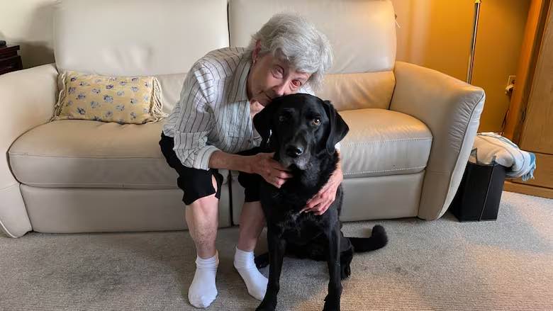
After 10 years without sight, Gail Lane, a 75-year-old woman from Victoria, B.C., has regained her vision. She can now see again thanks to a tooth surgically placed into her eye socket. Lane underwent osteo-odonto keratoprosthesis, a rare “tooth-in-eye” surgery, at Mount Saint Joseph Hospital in Vancouver. She was 1 of 3 Canadians to receive this rare surgery in February this year.
Lane lost her vision due to complications from Stevens-Johnson Syndrome, an autoimmune disorder usually triggered by certain medications. The complex two-part procedure helped her see her partner’s face and her dog’s wagging tail for the first time in 10 years. She is slowly beginning to be able to see light, then movement, and eventually facial features. The surgery uses a patient’s own tooth to anchor a plastic lens in the eye socket.
The Medical Journey Behind Corneal Blindness
Stevens Johnson Syndrome (SJS) severely damaged Lane’s corneas through scarring and inflammation. This rare autoimmune condition severely affects skin and mucous membranes, often triggered by certain medications like seizure drugs. The syndrome initially starts with flu-like symptoms followed by a painful rash that eventually spreads causing severe blistering. It also causes sloughing and scarring of the skin.
Corneal damage occurs in most cases of SJS and can lead to permanent blindness. Lane’s condition made conventional corneal transplants impossible due to severe ocular surface disease. Over 1.2 million Canadians live with vision loss which is about 3.2% of the population. The total cost of vision loss in Canada reached $32.9 billion in 2019.
Understanding Tooth-in-Eye Surgery Mechanics
Osteo-odonto keratoprosthesis involves extracting a patient’s tooth and implanting it with a plastic lens. Dr. Greg Moloney explained to the CBC “It’s a complex and strange operation, but it basically involves replacing the cornea”. The tooth forms a strong support structure for the plastic focusing telescope and the body readily accepts it without rejection.
The procedure uses the surrounding bone and the tooth of the patient to create support for the artificial cornea. Teeth contain dentin, which is the ideal tissue to hold a plastic lens with rejection from the body.
The Two-Stage Surgical Process
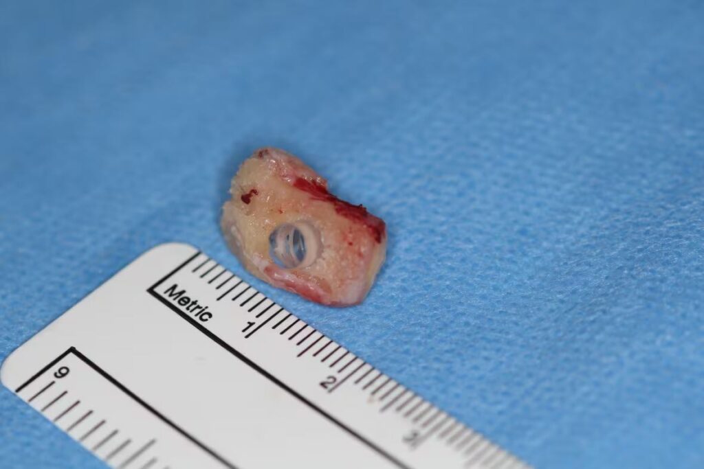
The operation is performed in 2 separate stages over several months. They first extract the tooth and implant it in the patient’s cheek. Then, they cover the eye with a layer of buccal mucous membrane to prepare the area.
After healing, specialists remove both the tooth and tissue from the cheek. They then insert a plastic lens into the tooth. Finally, the team sutures the tooth and lens into the eye socket. Each stage takes 6 to 8 hours and needs several experts. Healing between stages lets fresh tissue form around the tooth.
The Patient’s Remarkable Recovery Journey
Lane regained her vision gradually over several months after her February surgery. She first noticed light, then soon after, she saw movement and recognized her dog, Piper’s wagging tail. Over time, the black Labrador came into clear focus for her. Lane began to notice different parts of her surroundings more clearly.
Six months later, she finally saw her partner Phil’s face for the first time as they started their relationship after Lane had fallen blind. Now, she enjoys seeing vivid colors, trees, grass, and flowers. With prescription glasses, her vision reached 20/50, Dr. Moloney confirmed.
Historic First Canadian Procedures
Lane joined 2 other patients as Canada’s first tooth-in-eye surgery recipients in February 2025. Each patient underwent the procedure at Mount Saint Joseph Hospital. All 3 initial procedures proceeded without complications.
Dr. Moloney worked alongside Australian colleague Dr. Shannon Webber during these pioneering operations. Local surgeon Dr. Ben Kang received specialized training to continue the program. These surgeries mark a historic moment in Canadian medical innovation.
Success Rates and Long-Term Outcomes
OOKP surgery achieves remarkable long-term results worldwide. One 2022 Italian study reported a 94% anatomical survival rate over 30 years. About 60-70% of patients gained functional vision, reaching 6/18 acuity or better.
At 10 years, 66% of surgeries still survived, while 38% kept strong vision. Other studies recorded over 80% long-term retention at ten years. A systematic review found five-year anatomical survival averaged 87.8%, ranging from 67% to 100%. Experts agree OOKP provides the best long-term vision and device retention, especially for dry eye cases.
Managing Complications and Risks
Despite high success rates, tooth-in-eye procedures have significant surgical risks. Potential complications include infection, implant rejection, and tissue overgrowth. Glaucoma development is the most frequent complication of OOKP, affecting 7-47% of patients.
However, serious complications remain relatively rare with proper patient selection. Endophthalmitis occurs in less than 8% of cases. Retinal detachment rates vary between 0% and 26% depending on study populations. Most patients require ongoing specialized monitoring throughout their recovery. Lane experienced the procedures as “uncomfortable but not painful”.
The Future of Vision Restoration in Canada
The success of Canada’s first tooth-in-eye surgeries opens new possibilities for treating previously incurable blindness. Dr. Moloney hopes to establish Mount Saint Joseph as the national center for this procedure. Research continues into improved materials and techniques for keratoprosthesis surgery. Scientists explore alternatives to tooth tissue while maintaining biocompatibility advantages. As surgical techniques advance and accessibility improves, more people may experience the joy of seeing loved ones again.
Read More: This Type of Coffee May Raise Your Risk of Macular Degeneration By 700%