

The human eye is often described metaphorically as the “window to the soul,” but in clinical medicine, it is more accurately viewed as a non-invasive window into the human body’s circulatory and nervous systems. This unique anatomical arrangement allows doctors to observe live blood vessels, cranial nerves, and connective tissues in real-time, offering critical clues about a patient’s systemic health. Often, before a patient experiences generalized symptoms like fatigue, unexplained weight loss, or persistent pain, the delicate structures within the eye may already be signaling the presence of a chronic disease.
Understanding what your eyes say about your health is becoming a cornerstone of preventative medicine. A comprehensive dilated eye exam goes beyond simply checking visual acuity; it is a vital diagnostic tool that can reveal early signs of potentially life-threatening conditions. In recent years, groundbreaking research from scientific journals has validated the eye’s role in diagnosing complex diseases such as diabetes and various forms of cancer.
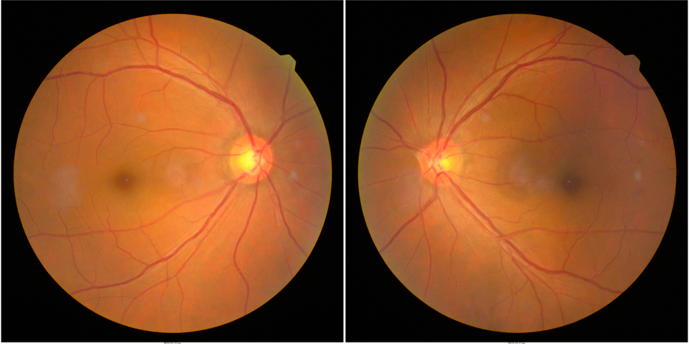
The Microvascular Signature of Diabetes

Image credit: Shutterstock
Diabetes mellitus is a metabolic disorder characterized by chronic hyperglycemia (high blood sugar). Over time, this elevated glucose damages the smallest blood vessels in the body, the capillaries, leading to widespread microvascular complications. Because the retina at the back of the eye is densely packed with these tiny vessels and has a high metabolic demand, it is one of the first areas to show damage. This condition is known as Diabetic Retinopathy (DR).
According to research published in journals such as The Lancet Diabetes & Endocrinology, diabetic retinopathy is a leading cause of vision loss globally, yet it is often asymptomatic in its earliest, most treatable stages. A doctor looking into your eyes can spot specific pathological changes that indicate diabetes is affecting your microvasculature.
The Progression of Damage: From Dots to Leakage
In the initial stages of DR (Non-proliferative Diabetic Retinopathy), the damage manifests as subtle, localized abnormalities. These are the following:
- Microaneurysms: These are often the earliest visible sign. Under fundoscopic examination, they appear as tiny red dots. Scientifically, they represent focal out-pouchings or balloons in the capillary walls caused by the loss of pericytes (cells that support vessel structure) due to high glucose stress.
- Hemorrhages: As the vessel walls continue to weaken, they may burst, leading to “dot-and-blot” hemorrhages within the retinal layers.
- Hard Exudates: Weakened vessels also become “leaky,” allowing lipids and proteins from the blood to seep into the retina. Doctors observe these as yellowish, waxy deposits with distinct borders.
The Proliferative Stage: A Pathological Response
If blood sugar remains uncontrolled, the retina suffers from widespread ischemia (lack of oxygen). In a pathological attempt to restore circulation, the body releases growth factors that stimulate Neovascularization, the growth of abnormal new blood vessels.
These new vessels are extremely fragile and poorly formed. They tend to leak severely into the vitreous (the clear gel filling the eye), causing sudden vision loss, and can pull on the retina, leading to retinal detachment. Spotting neovascularization is a critical signal of advanced, proliferative diabetes that requires immediate intervention.
Cancer: Primary Tumors, Metastasis, and Systemic Masquerade

Image credit: Shutterstock
While less common than diabetic retinopathy, the detection of cancer through an eye exam is a powerful diagnostic tool. The eyes can reveal cancer through three primary pathways: the presence of primary ocular tumors, the spread of metastatic cancer from another part of the body, or indirect systemic signs manifesting in ocular tissues.
Primary and Metastatic Ocular Tumors
The eye can be the site of origin for malignant tumors. The most common primary intraocular cancer in adults is Uveal Melanoma. This tumor arises from the melanocytes (pigment-producing cells) in the uveal tract (which includes the iris, ciliary body, and choroid). An ophthalmologist can detect this during a dilated exam as a pigmented (brown) or amelanotic (flesh-colored) mass beneath the retina. Early detection is vital, as uveal melanoma has a high potential to metastasize, particularly to the liver.
Conversely, because the eye, particularly the choroid layer, is highly vascular, it is a frequent site for Metastatic Cancer spreading from primary sites elsewhere in the body. According to oncology studies, the most common primary sources of choroidal metastasis are breast cancer in women and lung cancer in men. During an exam, these appear as creamy-white or yellowish, elevated lesions. In many cases, these ocular findings are the first indication that a patient has cancer elsewhere in their body.
Systemic Signals: Leukemia and Intracranial Pressure
Certain cancers, such as leukemia and lymphoma, are cancers of the blood or immune system, but they have distinct “masquerade: signals in the eye. For patients with leukemia, this presents as leukemic retinopathy. These patients often show signs of the disease in their retina even before they are diagnosed through a blood test. These signs include retinal hemorrhages with white centers, known as Roth spots, which represent bundles of leukemic cells or fibrin clotting. A doctor may also observe generalized swelling and tortuosity of the retinal veins.
Another sign of Papilledema, or optic nerve swelling. Brain tumors, whether primary or metastatic, can cause an increase in intracranial pressure, aka pressure inside the skull. This pressure is transmitted directly through the optic nerve sheath, which is connected to the brain. During a fundoscopic exam, this pressure causes the optic disc (where the optic nerve enters the eye) to swell and its margins to become blurred. Detecting papilledema is an emergency signal that requires immediate neurological imaging.
The Revolution of Oculomics: AI and Early Detection

The science of analyzing the eye to understand systemic health has given rise to a new, exciting medical field known as Oculomics. The core premise of oculomics is that the retina contains a wealth of structural and vascular biomarkers that can predict a person’s risk for systemic, cardiovascular, and neurodegenerative diseases.
The challenges have historically been the subtle nature of these early biomarkers. Many pathologically relevant changes are microscopic and escape detection by even the most experienced clinicians until the disease is advanced. This is where Artificial Intelligence (AI) and deep learning algorithms are revolutionizing diagnostics.
Super-Human Pattern Recognition
Several scientific journals have published seminal studies demonstrating that AI algorithms can be trained on massive datasets of hundreds of thousands of retinal images (fundus photographs and Optical Coherence Tomography (OCT) scans). By analyzing these images, AI can identify intricate patterns of microvascular narrowing, vessel tortuosity (twisting), branching angles, and texture changes that are invisible to the human eye.
With this comes many benefits. The first of these is early prediction. AI models have shown the ability to analyze a single retinal photograph and accurately predict a patient’s five-year risk of cardiovascular events, such as heart attack or stroke, with performance comparable to traditional risk factors like cholesterol levels and blood pressure.
AI is also now used in autonomous screening systems for diabetic retinopathy. These systems, such as IDx-DR (the first FDA-approved autonomous AI diagnostic system), analyze retinal images in a primary care setting and provide an immediate, reliable diagnostic decision without needing an ophthalmologist’s input. This increases access to screening and allows for earlier intervention.
Finally, AI also provides a window into Neurodegeneration. Oculomics is providing breakthrough insights into neurodegenerative diseases. Because the retina is embryonic brain tissue, the thinning of specific retinal layers (detected using OCT) can signal the early stages of Alzheimer’s and Parkinson’s disease, potentially years before cognitive symptoms appear.
Beyond Diabetes and Cancer: Other Illnesses Seen in the Eyes
The microvascular and neurological connectivity of the eye makes it sensitive to a wide array of systemic conditions. A routine dilated eye exam can lead to the incidental diagnosis of several other critical illnesses.
Hypertension (High Blood Pressure)
Like diabetes, hypertension primarily damages blood vessels. A doctor examining the retina can identify hypertensive retinopathy. Signs include “AV nicking”, where a stiffened artery compresses a vein where they cross, generalized narrowing of the retinal arteries, known as “silver” or “copper” wiring, and retinal hemorrhages. The severity of retinal damage often correlates directly with the risk of stroke and kidney damage.
Autoimmune and Inflammatory Diseases
Many systemic autoimmune diseases, such as Rheumatoid Arthritis, Lupus, Sarcoidosis, and Multiple Sclerosis (MS), cause generalized inflammation that can manifest in the eye. They can cause Uveitis, or inflammation of the uvea, the middle layer of the eye. While uveitis can have many causes, a significant number of cases are associated with underlying systemic inflammatory disorders. Another is optic neuritis, or inflammation of the optic nerve. This can cause sudden vision loss and pain. It is a common presenting symptom of Multiple Sclerosis.
Cardiovascular and Blood Disorders
The genetic blood disorder, sickle cell disease, can cause obstructions in the small vessels of the retina, leading to characteristic “sea-fan” neovascularization, aka abnormal vessel growth resembling a sea fan. If you have high cholesterol, doctors may observe Arcus Senilis, a white, gray, or blue ring around the cornea (the clear front window of the eye), caused by lipid deposits. While common in older adults, its presence in younger individuals can indicate dangerously high cholesterol. Additionally, tiny cholesterol emboli (Hollenhorst plaques) can sometimes be seen lodged within retinal arteries, signaling a significant risk of stroke.
The Bottom Line

Image credit: Shutterstock
The unique anatomy of the human eye provides a powerful, non-invasive diagnostic opportunity that is unrivaled in any other organ system. By directly visualizing the live blood vessels and nerves of the retina, clinicians can interpret delicate microvascular and neurological changes as a reflection of the patient’s generalized health. The science of what your eyes say about your health has been validated by decades of peer-reviewed research, cementing the dilated eye exam’s role as a critical component of preventative medical care.
From the specific microvascular signature of diabetic retinopathy to the complex ocular manifestations of primary and metastatic cancer, the eyes offer clues that can lead to early, life-saving interventions. Furthermore, the advent of AI-driven oculomics is unlocking new frontiers, transforming retinal imaging into a tool that cannot only detect existing disease but also predict a patient’s future risk for heart attack, stroke, and neurodegeneration.
Given that many of these systemic conditions are asymptomatic in their initial, most treatable stages, the message you need to know is clear: comprehensive, regular dilated eye exams are essential not just for preserving vision, but for safeguarding systemic well-being. By paying attention to what the eyes are saying, both patients and physicians can move from a model of reactive treatment to one of proactive, preventative medicine.
A.I. Disclaimer: This article was created with AI assistance and edited by a human for accuracy and clarity.
Read More: Bats May Hold a Key to Future Diabetes Therapies, Scientists Say