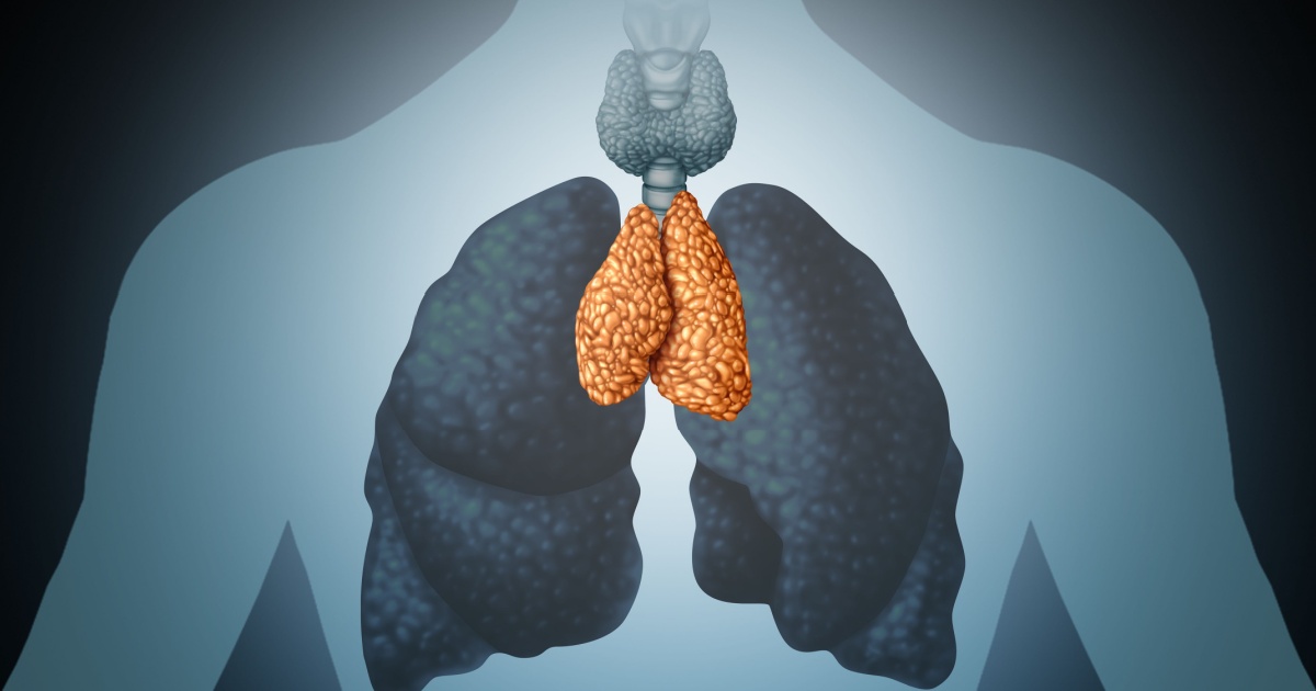

There is a small organ sitting behind your breastbone, roughly the size of a walnut in adults, that most physicians have spent decades barely thinking about. Medical students learn that it matters intensely in childhood, training the immune system’s T cells, and then gradually fades into irrelevance as the body matures. By the time most of us reach our thirties or forties, the prevailing assumption in clinical medicine has been that the thymus is essentially retired: shrunken, fatty, and functionally finished.
That assumption may now need to be retired instead.
Two studies from investigators at Mass General Brigham challenge a decades-old assumption that the thymus becomes irrelevant in adulthood. Using artificial intelligence to analyze routine CT scans, researchers uncovered that adults with a healthy thymus had increased longevity and reduced risk for cardiovascular disease and cancer, and in a separate study found that thymic health may influence response to immunotherapy. These findings, published in two papers in the same issue of Nature, suggest the thymus plays a far more consequential role in adult health than previously understood, and might provide a new target for personalizing disease prevention and cancer treatments.
We’ll take a look at both studies in detail: how the research was conducted, what it found, where its limits lie, and what it might mean for how adults think about their own immune health.
The Thymus: What It Does and Why Doctors Stopped Paying Attention
The thymus is a small organ that sits behind the breastbone. It is the only organ in the body capable of producing T cells. Those T cells are the immune system’s precision instruments – they identify and destroy infected cells, cancer cells, and other threats that the body needs to distinguish from healthy tissue.
The thymus is essential for establishing T cell diversity early in life, but undergoes profound involution with age and has therefore traditionally been regarded as largely nonfunctional in adults. “Involution” here means a physical and functional shrinking: the active thymic tissue is progressively replaced by fat, and the organ’s output of newly trained T cells falls sharply. With age, the glandular tissue in the thymus is replaced by fat, and the rate at which this happens is linked to sex, age, and lifestyle factors – findings that also indicate the appearance of the thymus reflects the aging of the immune system.
The downstream consequences of this process are significant. Thymic involution is a core characteristic of immunosenescence, characterized by gradual shrinkage of the thymus and a significant reduction in thymic epithelial tissue, leading to a pronounced decline in the production of T cells, particularly naïve T cells. As the thymus shrinks, T cell receptor diversity also decreases, impairing the ability of the immune system to respond effectively to novel pathogens. In practical terms: the immune system keeps its cell counts up, but does so by repeatedly copying a narrowing repertoire of older T cells. The immune library becomes smaller, less diverse, and less capable of mounting responses to genuinely new threats.
Immune system aging is characterized by the paradox of immunosenescence, literally “immune aging,” which describes disruption in the structural architecture of immune organs and dysfunction in immune responses, resulting from both aged innate and adaptive immunity.
For decades, doctors believed the organ was mostly inactive after puberty because it shrinks with age and produces fewer new T cells. As a result, its role in adult health has rarely been examined in large populations. The March 2026 studies are a direct challenge to that omission.
Study One: Thymic Health and Long-Term Health
In the first study, published in March 2026, researchers developed a deep learning framework to quantify thymic health from routine radiographic images and evaluated its association with longevity and risk of major age-associated diseases in two large prospective cohorts of asymptomatic adults: the National Lung Screening Trial (n = 25,031) and the Framingham Heart Study (n = 2,581).
The deep learning model analyzed CT scans – the kind routinely produced during lung cancer screening or cardiovascular assessment – and assessed each thymus for its size, shape, and tissue composition. The team analyzed the size, shape, and composition of the thymus, generating a “thymic health” score. Participants were then sorted into low, average, and high thymic health categories based on where their scores fell within the population distribution.
This is a critical methodological point. The researchers did not require any new scans or additional procedures. They extracted thymic health information from imaging that participants had already undergone for other reasons, making this approach potentially scalable to routine clinical practice in the future, if validated. In both cohorts, thymic health varied markedly across the population.
Survival Outcomes
The mortality findings were stark. People with high thymic health scores had about a 50% lower risk of death, 63% lower risk of cardiovascular death, and 36% lower risk of developing lung cancer compared to those with low thymic health. These associations remained significant after adjusting for age and other health factors.
In the National Lung Screening Trial, higher thymic health was consistently associated with lower all-cause mortality, reduced lung cancer incidence, and lower cardiovascular mortality over 12 years of follow-up, after adjustment for age, sex, smoking, and comorbidities.
The two new studies relied on scans and other data collected during the National Lung Screening Trial and participants in the ongoing Framingham Heart Study. The Framingham cohort, one of the longest-running cardiovascular population studies in the world, provided independent replication. Higher thymic health was significantly associated with reduced cardiovascular mortality in that cohort as well, independent of age, sex, and smoking.
Cancer Risk and Lung Cancer Incidence
The analysis showed that individuals with higher thymic functionality were less likely to develop lung cancer, with a 3.4% incidence at six years versus 5.3% for those with low scores. This association persisted even after accounting for smoking history, which is the dominant lung cancer risk factor, suggesting that thymic health may contribute to cancer surveillance capacity in a manner that is at least partially independent of tobacco exposure.
The researchers theorize that when thymic health and T cell diversity decline, the immune system may become less able to respond to new threats, like cancer or other diseases.
The Mechanism: Inflammation and Lifestyle
One of the most clinically significant aspects of the study was the identification of modifiable contributors to poor thymic health. Their analysis found that chronic inflammation, smoking, and high body weight were associated with poorer thymic health, suggesting that lifestyle and systemic inflammation may influence immune resilience across the lifespan.
Prior research in immunology has established the biological plausibility of this connection. Lifestyle factors are often referred to as determinants of healthy aging, and there is experimental evidence that antioxidant-rich diet and exercise attenuate immunosenescence and thymic atrophy. A 2023 study published in Immunity & Ageing on a Swedish middle-aged population found that former smoking, sedentary behavior, and low intake of vitamins, minerals, and fiber were all correlated with thymic scores in univariate analyses. The findings support the intriguing concept that obesity as well as low fiber intake contribute to immunological aging, thereby raising the possibility of preventive strategies.
These findings suggest a profound impact of actionable lifestyle choices on thymic health. They also clarify why healthy behavior improves well-being and lifespan in ways that extend beyond individual organ systems.
Study Two: Thymic Health as a Predictor of Immunotherapy Outcomes in Cancer Patients
While immunotherapy has changed cancer treatment profoundly, many patients still experience limited benefit, creating an urgent need for improved biomarkers to identify who will respond. Most existing biomarkers remain tumor-centric and largely overlook host immune competence.
Immunotherapy is a treatment that uses the body’s immune system, typically the patient’s own, to attack cancer. Immune checkpoint inhibitors – the class of drugs at the center of this second study – work by removing molecular “brakes” that cancer cells place on T cells, allowing those T cells to continue attacking tumors. The question the second Nature paper addressed was whether the underlying health of the thymus, the organ that trained and continues to supply those T cells, might determine how well patients respond.
Study Design and Findings
In the March 2026 paper, researchers applied a deep-learning framework to routine CT images to quantify thymic health in a pan-cancer cohort of 3,476 patients receiving immune checkpoint inhibitors.
Patients with stronger thymic health had about a 37% lower risk of cancer progression and a 44% lower risk of death, even after accounting for other patient, tumor, and treatment factors. These results came from Harvard-affiliated researchers at Brigham and Women’s Hospital and Dana-Farber Cancer Institute.
Among patients with non-small cell lung cancer, better thymic health was associated with lower risks for cancer progression and all-cause mortality, which was sustained with higher levels of PD-L1 expression and tumor mutation burden. PD-L1 expression and tumor mutation burden are the two biomarkers most commonly used today to predict who will benefit from checkpoint inhibitor therapy. The fact that thymic health’s predictive value persisted even when controlling for these established markers suggests it captures something distinct about the patient’s immune status – something tumor-focused tests cannot detect.
In an analysis of the prospective TRACERx lung cancer study, thymic health was positively associated with diversity of T-cell receptors and their excision circles, and also with immune-system signaling pathways. Findings were similar in patients with melanoma, breast cancer, and renal cancer.
This molecular validation is a crucial step. It demonstrates that what the AI “sees” on a CT scan corresponds to genuine, measurable biological activity – not merely anatomical appearance.
The Thymectomy Evidence: What Removal Has Already Taught Us
The 2026 Nature findings do not emerge in a vacuum. A pivotal study published in The New England Journal of Medicine had already provided indirect evidence for the adult thymus’s importance by studying what happens when it is surgically removed – a procedure called thymectomy (the-MECK-tuh-mee), which is routinely performed as part of certain cardiothoracic surgeries.
In the five years after surgeons removed their thymus, people were more than twice as likely to die of any cause compared to a similar group who had undergone heart or chest surgery but still had one. Those without thymuses were twice as likely to develop cancer.
These results strongly suggest that when possible, preservation of the thymus should be a clinical priority. The two studies now provide the population-level complement to that clinical evidence. Where the NEJM study showed what goes wrong when the thymus is removed, the new work shows that variation in thymic health across the general population, even in people who still have their thymus, tracks closely with the same outcomes: mortality, cancer, and cardiovascular disease.
Cellular Mechanisms: How the Thymus Ages and Why It Matters
The biology underpinning these findings has been clarified by parallel research into what actually happens inside the thymus as it ages. A 2024 study published in Nature Immunology identified specific cellular changes that drive thymic decline. Researchers applied single-cell and spatial transcriptomics, lineage-tracing, and advanced imaging to define age-related changes in nonhematopoietic stromal cells – the structural support cells of the thymus – and discovered the emergence of two atypical thymic epithelial cell states. These age-associated epithelial clusters formed dense regions devoid of developing thymocytes, representing an accretion of nonproductive thymic tissue that worsened with age.
Independent research published in 2024 in Nature Aging confirmed the downstream consequences: age-related thymic involution leads to a pronounced decline in the production of T cells, particularly naïve T cells, and as the thymus shrinks, T cell receptor diversity also decreases, impairing the ability of the immune system to respond effectively to novel pathogens.
What this cellular picture tells us is that thymic aging is not simply a matter of the organ getting smaller. The internal architecture becomes dysfunctional – it produces the wrong kinds of cells, loses its ability to repair damage, and contributes to broader systemic inflammation rather than dampening it.
Critical Limitations: What These Studies Cannot Yet Tell Us
Researchers noted that their findings need to be confirmed in future studies. They also propose looking at whether changing lifestyle factors might improve thymic function and if therapies like radiation might affect thymic health.
Several additional constraints deserve attention. The studies were observational, which means they can identify associations but cannot prove that poor thymic health causes premature death. It is possible that thymic decline and mortality share common upstream causes, such as chronic disease or poor metabolic health, rather than the thymus itself being the direct driver of outcomes. The sample in the primary cohort was also predominantly white, and future research in diverse populations is necessary before these findings can be generalized broadly.
The team is currently leading additional research to investigate whether other care-associated factors may impact thymic health. In one study, they are examining whether unintended radiation exposure to the thymus in patients with lung cancer may affect outcomes.
The imaging AI model provides valuable associations but is not yet validated as a diagnostic tool in routine medical practice. Longitudinal studies and clinical trials will be essential to confirm causality and determine whether interventions can reverse thymic decline or soften its health consequences.
Read More: 5 Ways to Keep Your Lymphatic System Healthy
What This Means for You
Together, these findings reposition the thymus as a central regulator of immune-mediated aging and disease susceptibility in adulthood, highlighting its potential as a target for preventive and regenerative strategies to promote healthy aging and longevity. That’s a significant claim, and it’s worth understanding what it does and doesn’t imply for anyone making everyday health decisions right now.
“The thymus has been overlooked for decades and may be a missing piece in explaining why people age differently, and why cancer treatments fail in some patients,” said Hugo Aerts, PhD, corresponding author on the papers and director of the Artificial Intelligence in Medicine Program at Mass General Brigham. For oncologists, the immunotherapy findings are especially relevant. Tumor-based biomarkers like PD-L1 expression and tumor mutation burden are currently the standard tools for selecting immunotherapy candidates. The finding that a host-based measure of thymic health predicts outcomes independently of those markers suggests that patient immune status deserves formal assessment in treatment planning – though that assessment tool is not yet ready for clinical deployment.
For everyone else, the most actionable takeaway lies in the lifestyle data. The modifiable factors linked to poorer thymic health – smoking, obesity, chronic inflammation, and physical inactivity – are the same ones targeted by existing public health recommendations. There is experimental evidence that antioxidant-rich diet and exercise attenuate immunosenescence and thymic atrophy. No special protocol or supplement is required to act on that information. Quitting smoking, maintaining a healthy weight, moving regularly, and eating a diet rich in fiber and micronutrients are all behaviors that existing evidence already supports – and that may, based on the patterns emerging from this research, support the longevity of the thymus as well. The 2026 Nature studies don’t change the recommendation. They add a compelling new reason to take it seriously.
Disclaimer: The author is not a licensed medical professional. The information provided is for general informational and educational purposes only and is based on research from publicly available, reputable sources. It is not intended to constitute, and should not be relied upon as, medical advice, diagnosis, or treatment. Always consult a licensed physician or other qualified healthcare provider regarding any medical condition, symptoms, or medications. Do not disregard, avoid, or delay seeking professional medical advice or treatment because of information contained herein.
AI Disclaimer: This article was created with the assistance of AI tools and reviewed by a human editor.
Read More: What You Need to Know About the Thymus: A Gland No One Talks About