

Dylan Jay Watts was living on the New South Wales South Coast when a violent chest infection overwhelmed him. According to Australian news reports, he died only days before turning 29, with scans revealing many cavities in his lungs. His sister Caitlyn stated that, “He was severely ill with a bacterium in his lungs that led to 72 holes that would never repair.” She went on to say that doctors had warned her that Dylan “would be lucky to make it to his 30th birthday.” Dylan’s Family and friends organized a fundraiser and told reporters that they were completely stunned by how quickly his condition had deteriorated. As Cheyenne O’Brien, a friend of the family, put it, “Although the 28-year-old had been gravely ill in the weeks and days leading to his passing, his loved ones were shocked by his death.”
From Chest Infection to Death

Local Australian news reported that the 28-year-old was the father of a young son and had passed away on the 15th of March. They noted that his lungs had been ravaged by a bacterial process that left a shocking number of holes. The holes were believed to be the result of a bacterial infection that had caused extensive destruction despite hospital treatment.
Dylan’s battles with his health started to worsen in 2021 after a scan indicated that he had two holes in his lungs. Halfway through 2024, the number of holes had surged to 72. In an interview with the Daily Australian Mail, his sister, Caitlyn, stated that “I found out in July of last year how severe things had begun to get, that he had holes in his lungs and had been in hospital for three weeks after coughing up blood.
His doctor told him if he hadn’t got in so soon, that could have killed him.” Dylan battled persistent symptoms, including a constant cough, reflux, overwhelming fatigue, and mounting mental health strain. When over-the-counter treatments brought no relief, a lung hemorrhage in early 2024 sent him to the hospital for three weeks, after which he began seeing specialists.
By that point, Dylan was on so much medication that it “filled an entire backpack.” In the days leading up to his death, his mother, Suzanne, brought Dylan and his puppy, Polar, home to set up long-term care. Sadly, Dylan passed away just five days later.
The Possible Cause of the Chest Infection
The pace of deterioration suggests a severe form of bacterial pneumonia, known to destroy tissue, called necrotizing pneumonia. In this variant of pneumonia, germs and the body’s immune response do not simply inflame air sacs; they kill and liquefy the lung tissue itself. A modern case review defined necrotizing pneumonia as the “main life-threatening manifestation of PVL SA infection, strongly linked with the need for ICU.” The initials “PVL SA” refer to Panton-Valentine leukocidin, a toxin made by some strains of Staphylococcus aureus.
Reports over the past two decades show that toxin-producing strains can cause rapidly progressive disease in young, otherwise healthy people. This is especially true when an influenza-like illness has recently worsened conditions. One early review summarized the pattern in very stark terms. Necrotizing pneumonia due to Panton-Valentine leukocidin-positive Staphylococcus aureus is “usually severe and often fatal, involving primarily young and healthy patients.”
You can think of a healthy lung as a fine sponge of delicate air sacs where oxygen crosses into the blood. In those with necrotizing pneumonia, portions of that sponge start to die. The dead tissue then breaks down and leaves air-filled spaces that radiologists call cavities. These cavities can then subsequently merge and grow. Blood vessels may be exposed and eroded, which explains why coughing up blood often becomes a frightening but common sign. Air then escapes from damaged tissue into the chest cavity, potentially causing the lung to collapse.
When bacteria enter the bloodstream, they can trigger a chain reaction in the body’s immune and clotting systems. This then leads to sepsis, dangerously low blood pressure, and even organ failure. At the molecular level, Panton-Valentine leukocidin creates pores in white blood cells, triggering inflammation and tissue damage.
How Bacteria Trigger Necrotizing Pneumonia

Several bacteria can trigger necrotizing pneumonia, and three in particular appear again and again in clinical series. These are Streptococcus pneumoniae, Staphylococcus aureus, and Klebsiella pneumoniae. Strains of Staphylococcus aureus that carry the genes for Panton-Valentine leukocidin are of particular concern. This is because these cases often move fastest and strike hardest in people who were not previously frail. Additionally, many studies have linked Panton-Valentine leukocidin-positive Staphylococcus aureus to an increased risk of intensive care admission and a potentially complicated recovery.
According to an MDPI review, severe cases should receive a regimen that includes an antitoxin agent alongside bactericidal antibiotics. The authors noted that, “The treatment of severe cases should include an anti toxin molecule and timely source control.” Studies have also noted that a preceding virus can make you more susceptible to future bacterial damage. So, was necrotizing pneumonia the cause of Dylan’s fatal chest infection?
Well, it is important to acknowledge that the press or family have not publicly stated the actual organism that caused the holes to appear in his lungs. Yet, they did mention early cavities that multiplied over months, recurrent hospitalizations, coughing blood, and a heavy mix of medicines. This is typically consistent with severe necrotizing bacterial pneumonia that resists any attempts to hinder or treat the process. It also fits with how clinicians experience a minority of pneumonia cases that turn into tissue damage rather than regular inflammation.
In such cases, the doctors will usually start by giving the patient broad-spectrum antibiotics while cultures are pending. After that, they will add agents that suppress toxin production if Staphylococcus aureus is suspected. The next step is to then drain away any collections of pus or air, and provide the patient with mechanical breathing support if needed.
Red Flags to Look Out for in Chest Infections
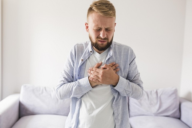
Most chest infections tend to improve, given enough rest, medicines, and time. However, there are those that do not. There are many situations where immediate medical evaluation of a chest infection is vital. This includes a sustained high fever, a feeling of air hunger, very fast breathing, a new sharp chest pain that worsens with each breath, confusion or faintness, or coughing up blood.
According to the Cleveland Clinic, pneumonia makes breathing difficult and can produce a cough with yellow, green, or bloody mucus. They go on to emphasize that treatment depends on how severe things are. They also advise seeking emergency care when breathing is difficult or chest pain is present because “pneumonia can get worse quickly.”
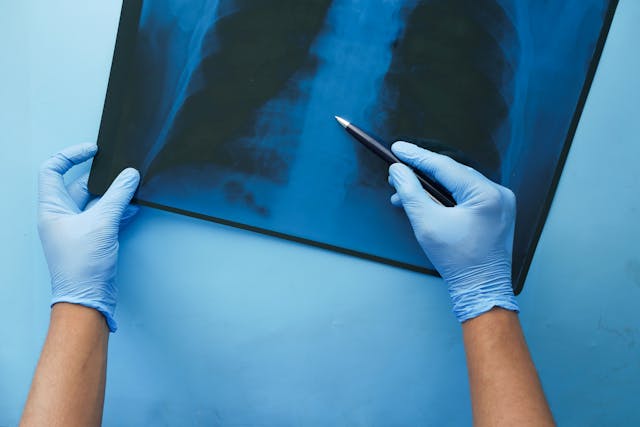
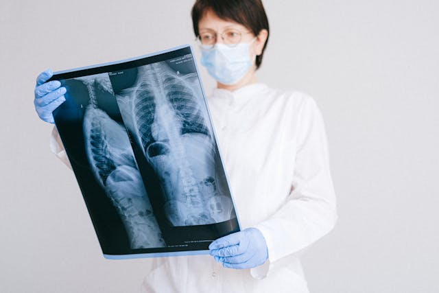
The clinical recommendations for front-line doctors offer the same advice: if a chest infection suddenly causes sharp, stabbing chest pain, it could be a collapsed lung on top of pneumonia, so the person needs urgent hospital care. Once a person with suspected necrotizing pneumonia reaches care, imaging and microbiology help guide their next moves. They usually use a chest X-ray for the initial examination. However, computed tomography is much better at showing the number and location of cavities and at spotting complications that may need drainage.
Additionally, blood work can reveal a high inflammatory response or, in toxin-associated cases, a paradoxically low white blood cell count. Various cultures taken from sputum and samples obtained through bronchoscopy can help identify the organism, so that they can pick the right antibiotics to use.
When Lung Tissue Starts to Break Down
Treating necrotizing pneumonia is a race to stay ahead of a very destructive process. Doctors first use broad antibiotics, then fully focus on the culprit once identified by the cultures. When breathing fails, the doctors typically deliver high-concentration oxygen or mechanical ventilation to the patient. They drain infected fluid around the lungs, and they evacuate air that has leaked into the chest cavity. Surgeons are usually very cautious during the active destructive phase because cutting into a sick lung can worsen air leaks and bleeding.
However, sometimes surgery is the only option when bleeding will not stop or tissue death is widespread. When several lobes are involved or when the illness begins after an influenza-like prodrome, hospital stays are often long, and patients don’t fully recover. Some reports describe survivors who improved after being given combinations that included clindamycin to suppress toxin formation alongside agents that kill the bacteria directly.
A common feature of many rapidly fatal cases is the appearance of bacterial necrotizing pneumonia after an influenza-like illness. The virus damages the airway lining and stunts local defenses, which allows the bacteria to gain a foothold. Various reports also reveal a common pattern: a person gets influenza B, which weakens the airways. After that, a toxin-producing Staphylococcus aureus infection moves in and causes severe pneumonia, even in people who were healthy before.
One medical report spoke of a healthy person who first got influenza B. Soon after, a strain of Staphylococcus aureus that makes powerful toxins took hold in their lungs and caused tissue-destroying pneumonia. This illustrates how an ordinary winter flu can weaken defenses and let a rare but very aggressive bacterial infection take over, even in someone who seemed well before.
Read More: Mother’s Sinus Infection Symptoms Turn Out to Be Cancer Diagnosis
The Reality of Long-Term Recovery for Survivors
Even when patients survive necrotizing pneumonia, the path back to normal life can be slow. Children often have stronger healing potential when they get sick, yet follow-up studies reveal lasting reductions in exercise capacity months after discharge. Adults who have lost large regions of lung tissue may experience a chronic cough and an increased vulnerability to future infections.
This is due to the way the disease damages the lungs. Once the sponge-like architecture of the lung is replaced by scars and cavities, the surface area for gas exchange is permanently reduced. Necrotizing pneumonia is uncommon compared with normal pneumonia, but when it occurs, it is typically associated with longer hospital stays and a higher risk of death or disability.
Not everyone is fortunate enough to survive, though, as was the case with Dylan. His sister Caitlyn wrote, “There is a fragility in all of us; his memory and his story will be remembered by many who loved him, and he will be forever missed.” She also encouraged others to act on the lesson of sudden loss. “Let this be a reminder that those you love can be gone in an instant, so hold them close today and tell them you love them,” she stated.
It is also a reminder not to ignore any signs of a serious chest infection or pneumonia. According to the Cleveland Clinic, pneumonia “makes it difficult to breathe and can cause a fever and cough with yellow, green, or bloody mucus.” If you have chest pain or difficulty breathing, seek emergency care as soon as possible because pneumonia can worsen quickly.
The Bottom Line
The precise organism in Dylan’s case has not yet been made public, and there is no verified final diagnosis in the open record. Yet, the pattern described by family and friends, and the language used by multiple outlets, sounds like necrotizing bacterial pneumonia that resisted therapy.
That framework explains the multiplication of cavities, the long hospital stays, the heavy medication load, and the devastating final turn. It also explains the doctors’ warnings that even if he stabilized, the holes would not simply close. Yet, stories like this one carry a human message beyond all the clinical details. Dylan’s sudden degradation and passing remind us all how truly fragile and short life is.
It reminds us that we need to be aware of how quickly those we love can be taken away from us. Dylan’s sister reminded us that we should take the time to tell those we love that we love them. However, there is also an important health message. Influenza vaccination reduces the risk of the viral stage that can open the door to a destructive secondary pneumonia. Timely medical assessment when severe symptoms appear gives clinicians a chance to identify a toxin-producing bacterium. They can then pair bactericidal and anti-toxin antibiotics while there is still a viable lung to save.
Disclaimer: This article was created with AI assistance and edited by a human for accuracy and clarity.
Read More: The Ability to Reverse Damage to Your Heart and Lungs is Closer Than You Think