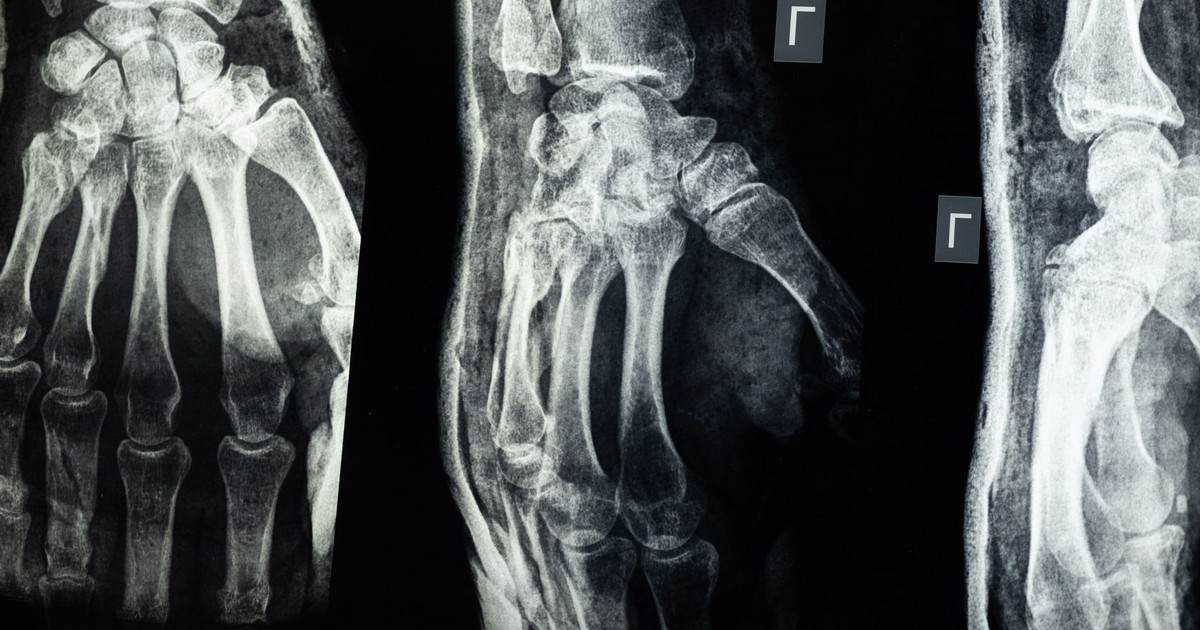

For millions of people with osteoporosis, treatment has traditionally focused on one major goal: slow down the loss of bone before another fracture happens.
But what if damaged bone could actually be rebuilt?
Researchers in Spain have taken an unusual step toward that possibility using patients’ own stem cells. The cells were removed, modified in a laboratory to help them find their way back to bone, and then returned to the body through a single intravenous infusion.
The first human trial was small, involving just 10 women with severe osteoporosis, and it was primarily designed to determine whether the treatment was safe.
But researchers saw something they weren’t necessarily looking for.
In the two years before treatment, the women had experienced an average of eight fractures per year as a group. During the two years afterward, that fell to just 0.5 per year. Bone samples and imaging also showed signs that new bone was being formed.
The results, published in Cell in September 2026, are far too early to show that the treatment prevents fractures. There was no placebo group, and only 10 people participated.
Still, the study raises an intriguing possibility: could a patient’s own cells eventually be used to help rebuild bone weakened by osteoporosis?
What osteoporosis actually does to bone
Osteoporosis is a gradual metabolic bone disease characterized by decreased bone mass and degradation of bone microarchitecture. Healthy bone is not solid like concrete; it has an internal lattice structure, and that structure determines how much force the bone can absorb before it breaks.
Available drug therapy for osteoporosis primarily targets the inhibition of bone resorption and agents that promote bone mineralization, designed to slow disease progression. Safe and predictable means to increase bone formation have been elusive. Bisphosphonates, the most widely prescribed class of drugs for the condition, work by suppressing osteoclasts, the cells that break down old bone tissue. They do not build new bone; they slow the rate of loss. Extended use of these medications has drawn attention to two severe, if rare, adverse events: atypical femoral fracture and medication-related osteonecrosis of the jaw, which are more common in patients with high cumulative doses and longer duration of therapy.
Current pharmacological treatments, such as bisphosphonates, selective estrogen receptor modulators, and anabolic agents, can reduce fracture risk; however, their prolonged use is limited by significant adverse effects, elevated treatment costs, and a lack of sustained disease remission. Regenerative medicine is now trying to address that gap between managing decline and actually reversing it.
According to the International Osteoporosis Foundation, approximately 500 million men and women worldwide may be affected by the condition, with up to 37 million fragility fractures occurring annually in individuals aged over 55. One in three women and one in five men aged over 50 will experience osteoporotic fractures in their lifetime. A fracture from osteoporosis often marks the beginning of a cycle of hospitalizations, lost mobility, and, for older patients, significantly elevated mortality risk.
Why bone loses ground in the first place
Bone remodeling relies on the coordinated activity of osteoclasts, which resorb old or damaged bone, and osteoblasts, which build new bone tissue. In a healthy adult, this process maintains a rough equilibrium. An imbalance between bone resorption and bone formation can cause bone diseases like osteoporosis, where the destruction side of the equation consistently outpaces the building side.
Osteoblasts, the cells responsible for laying down new bone, originate from mesenchymal stem cells (MSCs). These bone marrow mesenchymal stem cells can differentiate in the laboratory into osteoblasts, chondrocytes, and adipocytes – meaning they can become bone-forming cells, cartilage cells, or fat cells depending on what signals they receive.
People with osteoporosis may have enough of these stem cells, but too few of them successfully reach the bone marrow and complete the job of forming new bone. MSCs reside primarily in the bone marrow but also exist in other sites such as adipose tissue. When they are extracted, cultured in the lab, and reintroduced through the bloodstream, they face a navigation problem.
The molecule that was missing
The bone marrow does not accept cells indiscriminately. Cells traveling through the bloodstream use specific surface molecules to identify and bind to the correct tissue. When MSCs are grown outside the body, they lose a surface molecule called Sialylated Lewis X, known as sLeX. Without sLeX, conventionally cultured MSCs infused intravenously tend to miss their bone marrow target.
The research team, which included hematologist José M. Moraleda of the University of Murcia in Spain, addressed this using a technique called glycocalyx editing. The glycocalyx is a sugar-based coating on the surface of cells that governs how they interact with their surroundings. The MSCs were modified to restore sLeX expression, improving their ability to return to the bone marrow after reintroduction to the bloodstream. This modification reprogrammed the cells to find and settle in bone tissue – a property the researchers called osteotropism, meaning bone-seeking behavior.
Prior animal studies had confirmed this approach could work in preclinical models. The University of Murcia trial was designed to determine whether the same modification would be safe and effective in human patients.
What the trial did and what it found
In the small trial involving 10 women with advanced osteoporosis aged between 51 and 72, the researchers extracted bone marrow from each participant’s hip. The MSCs were then isolated and grown in the laboratory, modified to restore sLeX expression, and returned to each patient through a single intravenous infusion. Participants were then monitored across approximately six years of follow-up.
On safety, the results were unambiguous. The experimental treatment was well tolerated by the participants, with no treatment-related adverse events occurring. The biological evidence also pointed to actual bone regeneration. Bone tissue biopsies taken 120 days after the MSC infusion showed a significant increase in average bone tissue area for seven of the 10 patients.
Bone metabolism biomarkers – proteins in the blood that signal new bone formation – also shifted in a direction consistent with increased bone-building activity. Imaging of the skeleton showed improvements in trabecular bone density, which is the spongy, lattice-like inner bone that osteoporosis attacks first.
Trabecular bone is metabolically active, making it particularly sensitive to hormonal and mechanical influences, and it is also the type of bone most vulnerable to early-stage osteoporosis. Bone loss in early osteoporosis is mainly trabecular, and with increasing age becomes primarily endocortical and intracortical. The trial showed regenerative effects concentrated in trabecular bone, meaning the treatment appears to be acting where the disease hits hardest and earliest.
The fracture numbers are the story
In the two years before their infusions, the 10 women in the trial experienced a combined rate of eight fracture events per year. In the two years after, that rate fell to 0.5 events per year. The researchers describe the drop as “precipitous,” and contextually, it is; these were patients already classified at very high fracture risk. Protecting them from recurrent breaks using a single intervention, if that finding holds in larger studies, would represent a significant departure from the chronic disease management model that defines current osteoporosis care.
Clinically, “very high risk” means documented, recurrent fractures have already occurred. Many such fractures from osteoporosis go unrecognized in standard medical settings, even when they happen. As noted in an earlier Hearty Soul piece on bone fractures, two-thirds of spinal fractures in the US go undiagnosed each year, often because they cause no immediate pain. Patients at very high risk have already experienced documented breaks, meaning the stakes of any new treatment are high.
What the research cannot yet claim
Researchers on the trial are explicit about the limits of what this study demonstrates. It was a phase 1 safety study, not a randomized controlled trial. There was no placebo group. There was no comparison arm of patients receiving standard care. The 10 participants were all women, leaving questions about how the approach might perform in men or in younger patients. The trial was conducted at a single center, and the patient pool lacked demographic diversity.
This trial is one of the most advanced human data points in the restorative direction, but it remains an early data point. Larger multi-site trials with randomization and control groups are required before any clinical conclusions can be drawn about efficacy. The researchers acknowledge this directly in their paper.
The modified MSC approach appears safe at this scale, it is technically feasible to extract, culture, modify, and re-infuse a patient’s own stem cells, and the biological signals in bone tissue moved in the right direction in most participants.
What happens now?
For now, this isn’t a treatment someone with osteoporosis can ask their doctor for. Ten patients in an early safety trial are nowhere near enough to establish that modified stem cells can reliably prevent fractures, and larger controlled trials will be needed.
But that’s also what makes the results interesting.
Researchers weren’t simply trying another way to slow the breakdown of bone. They modified patients’ own cells so those cells could better find their way back to bone tissue, where the study found signs of new bone formation.
The dramatic drop in fractures is especially intriguing, but without a control group, researchers cannot yet say the stem cell treatment caused it. That question will have to be answered by much larger studies.
Current osteoporosis treatments remain the standard of care and have strong evidence showing they can reduce fracture risk. This experimental approach isn’t ready to replace them.
Instead, it points toward a very different possibility for the future of osteoporosis treatment.
Rather than only trying to stop bones from getting weaker, researchers are beginning to ask whether they can help the body build them back.
Disclaimer: The author is not a licensed medical professional. The information provided is for general informational and educational purposes only and is based on research from publicly available, reputable sources. It is not intended to constitute, and should not be relied upon as, medical advice, diagnosis, or treatment. Always consult a licensed physician or other qualified healthcare provider regarding any medical condition, symptoms, or medications. Do not disregard, avoid, or delay seeking professional medical advice or treatment because of information contained herein.
AI Disclaimer: This article was created with the assistance of AI tools and reviewed by a human editor.
Read More: Scientists May Have Been Wrong About How ADHD Medications Work