

One of the most precious bonding moments for expecting mothers is when they get to see an ultrasound of their unborn child for the first time. Many sight-impaired mothers are unable to experience this, until relatively recently. With 3D ultrasound technology, blind mothers can finally have a “look” at their unborn children.
3D Ultrasound Allows Blind Mother to See Her Unborn Child

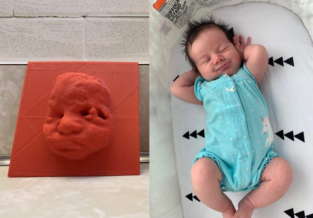
An emotional video posted by Huggies Brazil on YouTube shows the moment when a blind mother gets to “see” her unborn son for the first time. This was all thanks to 3D ultrasound printing technology. (1) The video shows 30-year-old Tatiana Guerra, who has been blind since she was 17, arriving for her ultrasound appointment. The ultrasound technician begins by describing what he sees on the screen of her 20-week-old fetus. The 3D ultrasound then prints a relief sculpture of her son Murilo’s face. Tatiana traces her fingers over the child’s face, crying tears of joy at “seeing” her baby for the first time. (1)
Another Happy Story
A mother from Baltimore also got to experience the joy of seeing her baby’s ultrasound thanks to 3D ultrasound technology. (4) Born with glaucoma, Taylor Ellis had little vision her whole life. Her sight was strong enough to see her first two children’s ultrasound images, but this time was different. When she went in for her 20-week ultrasound, she was devastated to discover that she no longer had enough sight to see her child’s photo. (4)
Her doctors knew how upset she was, so they gave her a special 3D ultrasound that printed her unborn baby’s face. This gift wasn’t just for her, either. Taylor’s husband Jeremy is also visually impaired, so he, too, was able to “see” their child for the first time. (4) Usually, John Hopkins Hospital uses this technology to print 3D models of babies with spina bifida, so that the surgeons can learn whether or not they need to do in-utero surgery. (4) For both Taylor and Jeremy, 3D ultrasound technology has been a dream come true. (4)
A Bumpy Start of 3D Ultrasound Tech

3D ultrasound technology first made an appearance in 2012 in Japan. It was as a way for expectant parents to have a keepsake figure of their unborn fetus. It quickly spread to the U.S., South Korea, and Estonia, where parents could pay up to $1,300 for one of these figurines. (2) Dubbed “weird fetus dolls” by much of the internet, these 3D ultrasounds weren’t really viewed positively until the video of Tatiana appeared. (2) The Huggies Brazil video was viewed over 1.6 million times and has certainly opened people’s eyes to the benefits of 3D ultrasound technology. (2)
Different Types of Ultrasounds

Today there are many options for ultrasounds, including (3):
- 2D
- 3D
- 4D
- Doppler
It can be confusing learning the differences between them all and most importantly when you should be having them. Here is a quick breakdown for you. (3)
Traditional 2D Ultrasound

This is the exam that most women are used to having to see their baby for the first time. In this ultrasound, a wand is passed either over the belly or into the vagina to send sound waves throughout the body. The waves bounce off of the internal organs and fluids, and their “echoes” are translated by a computer into the image of the fetus that you see on the screen. (3)
Read: My Husband’s Reaction to Finding Out Our Baby Girl Has No Arms or Hands Put the Doctors to Shame
Doppler Ultrasound

In this ultrasound, the technician uses a hand-held ultrasound device and a special jelly on the mother’s belly to amplify the sound of the fetal heartbeat. (3)
3D Ultrasound

To achieve these, technicians take multiple 2D images from a variety of angles which are then put together to form a 3D rendering of the fetus. (3)
4D Ultrasound
https://www.pexels.com/photo/photo-of-ultrasound-testing-7088840
Similar to the 3D except it is more like a video and shows movement. 4D ultrasounds allow parents to see their babies moving in real time. (3) Usually, 3D and 4D ultrasounds are used to examine abnormalities and to monitor specific conditions, such as cleft lip or spinal cord issues. They are beginning to promote novel services where parents can have keepsake portraits of their children, however, the American Congress of Obstetricians and Gynecologists (ACOG) and the FDA warn against expecting mothers having them done. (3) The ACOG and FDA agree that while there are no risks from ultrasounds for pregnant women, they should still be used carefully and only for medical purposes by a qualified professional when deemed medically necessary. (3)
Current Recommendations
https://www.pexels.com/photo/people-woman-technology-computer-7089015
The current recommendation by the ACOG is that every expecting mother should have at least one 2D ultrasound between 18 and 22 weeks of pregnancy. (3) They discourage using all other forms, including 3D ultrasounds, for the purpose of a memento because (3):
- You potentially put your baby’s health at risk
- Commercial ultrasound technicians likely do not have the knowledge and expertise to answer questions or spot developmental abnormalities
- The sessions are long (up to 45 minutes) which is intrusive and disruptive for a growing fetus
As always, ask your doctor before you take any medical advice or otherwise from third parties. The health of you and your baby is the most important thing during pregnancy – there will be plenty of times to take pictures of your baby after they are born.
Sources
- https://www.cbsnews.com/news/3d-printed-ultrasound-lets-blind-mom-see-unborn-baby/#:~:text=A%20video%20posted%20on%20YouTube,at%20the%20age%20of%2017
- https://www.cbc.ca/news/trending/3d-printed-ultrasound-lets-blind-woman-see-her-unborn-baby-1.3065489
- https://www.whattoexpect.com/pregnancy/pregnancy-health/prenatal-testing-3d-4d-ultrasounds/
- https://www.goodnewsnetwork.org/blind-mom-sees-baby-through-3d-ultrasound/